ECG
Diterbitkan
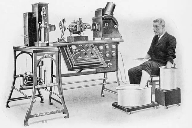
Willem Einthoven, father of ECG, Nobel Prize 1924
Oleh
Zarif Fathurrahman Rani
5 minit baca
ECG
Introduction
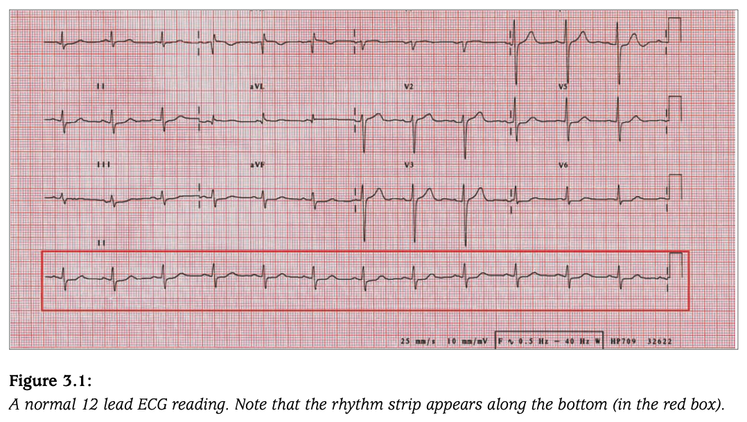
Electrocardiogram (ECG) is a way of reading electrical movement of the heart. Electrical impulse started from Sinoatrial node (SA node) causing atrial contraction. Move to Atrioventricular node (AV node), Bundle of His then Bundle Branch and end at Purkinje fiber in which cause ventricular contraction. In this blog, i will present a systematic approach to reading an ECG. Knowledge and illustration from this blog was extracted from The Ecg Workbook (Angela Rowlands, Andrew Sargent), The ECG Made Easy 8th edition and Live In The Fastlane (LITFL) website
Flow of reading
- Rhythm strip
- Limb lead
- Chest lead
- Heart injury
- Bundle branch block
- Chamber enlargement
Rhythm Strip
 Rhythm strip is the Lead ll in the ECG
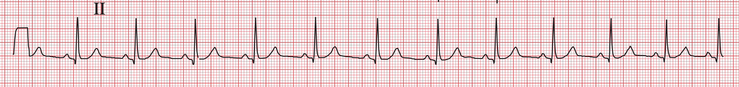 Normal sinus rhythm
Rate
- Normal: 60 - 100 bpm
- Arrhythmia: Tachycardia > 100, Bradycardia < 60
- Calculate peak R wave in 6 seconds / 30 large box
Rhythm
- Normal: regular rhythm / equal distance between peak R wave
- Abnormal: Regularly irregular, Irregularly irregular
P wave
- Normal: present, < 120 msec
- Abnormal: absent / inverted / prolong / P-mitral / P-pulmonale
QRS complex
- Normal: narrow, < 120 msec
- Abnormal: broad / prolong
PR interval
- Normal: 120 - 200 msec
- Abnormal: < 120 msec / > 200 msec
P:QRS ratio
- Normal: 1:1
- Abnormal: 1:0 / 3:1 / others
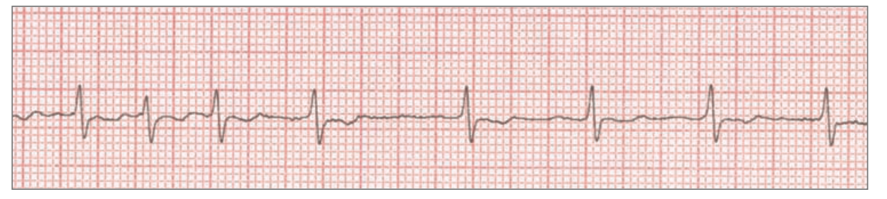
Arrhythmia
- Sinus origin / physiological
- Complete visualization of PQRST complex with normal morphology
- Sinus tachycardia / sinus bradycardia
- Atrial fibrillation
- Most common cardiac arrhythmia
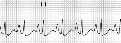
- ECG finding: tachycardia, irregularly irregular, absent P wave, narrow QRS
 ~150 bpm irregularly irregular, no discrete P wave, narrow QRS, N/A PR, N/A P:QRS, (irregular irregular tachycardia with no discrete P wave and narrow QRS) *Atrial Fibrillation
- Atrial flutter
- ECG finding: tachycardia, broad multi P wave / F wave, P:QRS ratio is 2:1 / 3:1, narrow QRS
 ~100bpm regular, broad multi P wave / F wave / saw tooth, narrow QRS, N/A PR, P:QRS 3:1, (saw tooth appearance with P:QRS ratio of 3:1) *Atrial flutter
- ECG finding: tachycardia, broad multi P wave / F wave, P:QRS ratio is 2:1 / 3:1, narrow QRS
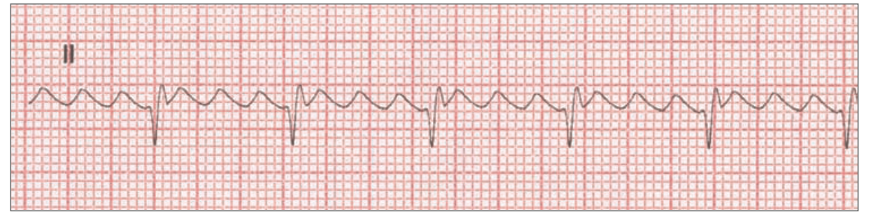
- Supraventricular tachycardia *AV node origin
- ECG finding: tachycardia, absent P wave, narrow QRS complex
 ~150bpm regular, absent P wave, narrow QRS, N/A PR, N/A P:QRS (tachycardia with narrow QRS) *supraventricular tachycardia
- ECG finding: tachycardia, absent P wave, narrow QRS complex
- Ventricular tachycardia *Ventricular node origin
- ECG finding: tachycardia, absent P wave, bizarre broad QRS complex
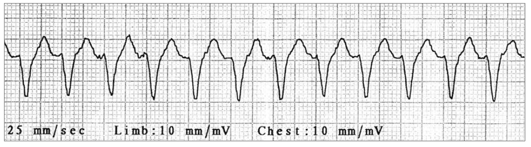 ~150bpm regular, absent P wave, broad QRS, n/a PR, n/a P:QRS, (tachycardia with broad QRS) *ventricular tachycardia
- ECG finding: tachycardia, absent P wave, bizarre broad QRS complex
- Ventricular fibrillation
- ECG finding: no P wave, no QRS complex, chaotic activity
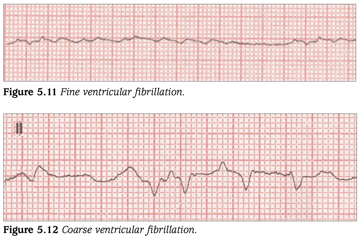 no rate and rhythm, no P wave, no QRS, n/a PR, n/a P:QRS
- ECG finding: no P wave, no QRS complex, chaotic activity
Heart Block
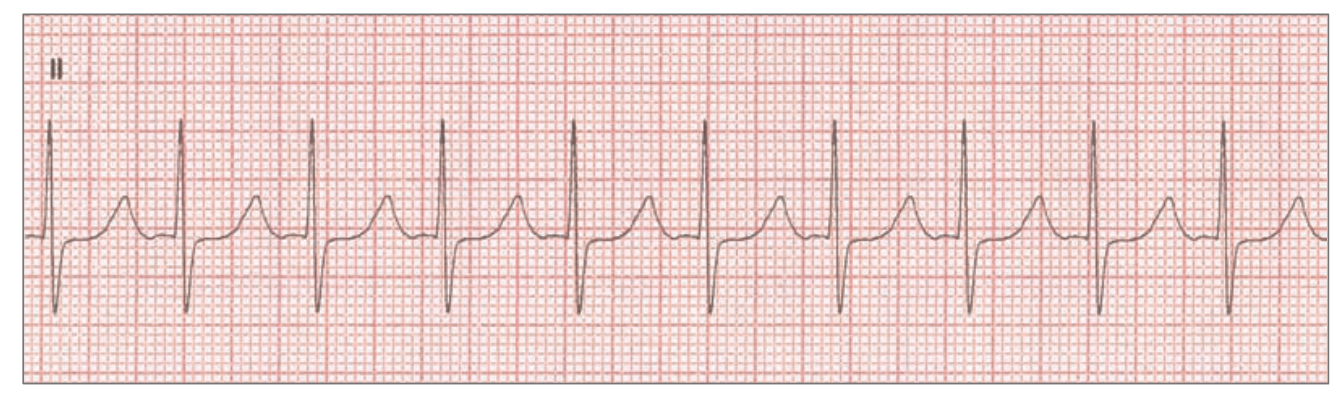
- 1st degree heart block (QRS complex present)
- ECG finding: PR < 200 msec, others normal
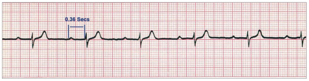 PR interval 360 msec
- ECG finding: PR < 200 msec, others normal
- 2nd degree heart block (QRS complex absent)
- Mobitz type l (Wenckebach phenomenon)
- ECG finding: PR interval progressively prolong until missing a QRS complex
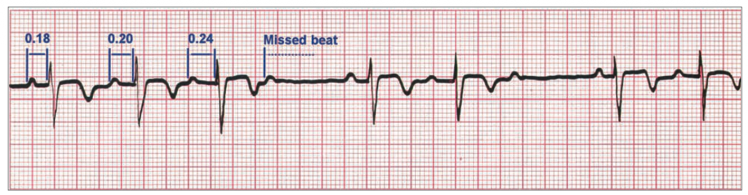 ~60bpm irregular ventricular rhythm regular atrial rhythm , normal P wave, narrow QRS, PR interval progressively prolong until missing a QRS, *second degree heart block - Mobitz type l
- ECG finding: PR interval progressively prolong until missing a QRS complex
- Mobitz type ll
- ECG finding: PR interval constant with intermittent absent QRS complex, P:QRS not 1:1 ratio,
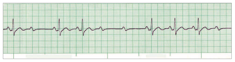 ~60bpm irregular ventricular rhythm regular atrial rhythms, normal P waves, narrow QRS, constantly prolong PR, P:QRS intermittent absent QRS *second degree heart block - Mobitz type ll
- ECG finding: PR interval constant with intermittent absent QRS complex, P:QRS not 1:1 ratio,
- Mobitz type l (Wenckebach phenomenon)
- 3rd degree heart block (Complete heart block)
- ECG finding: P:QRS independent, broad QRS
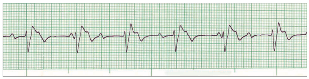 ~60bpm regular, intermittent absent P wave, broad QRS, prolong PR, P:QRS independent *third degree heart block
- ECG finding: P:QRS independent, broad QRS
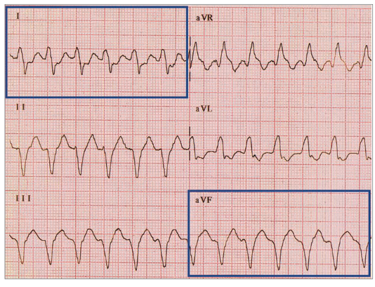
Limb Lead
 Limb lead ECG
Cardiac Axis
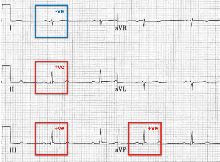
- Normal cardiac axis
- ECG finding: Lead l and aVF being positive
 Normal axis deviation
- ECG finding: Lead l and aVF being positive
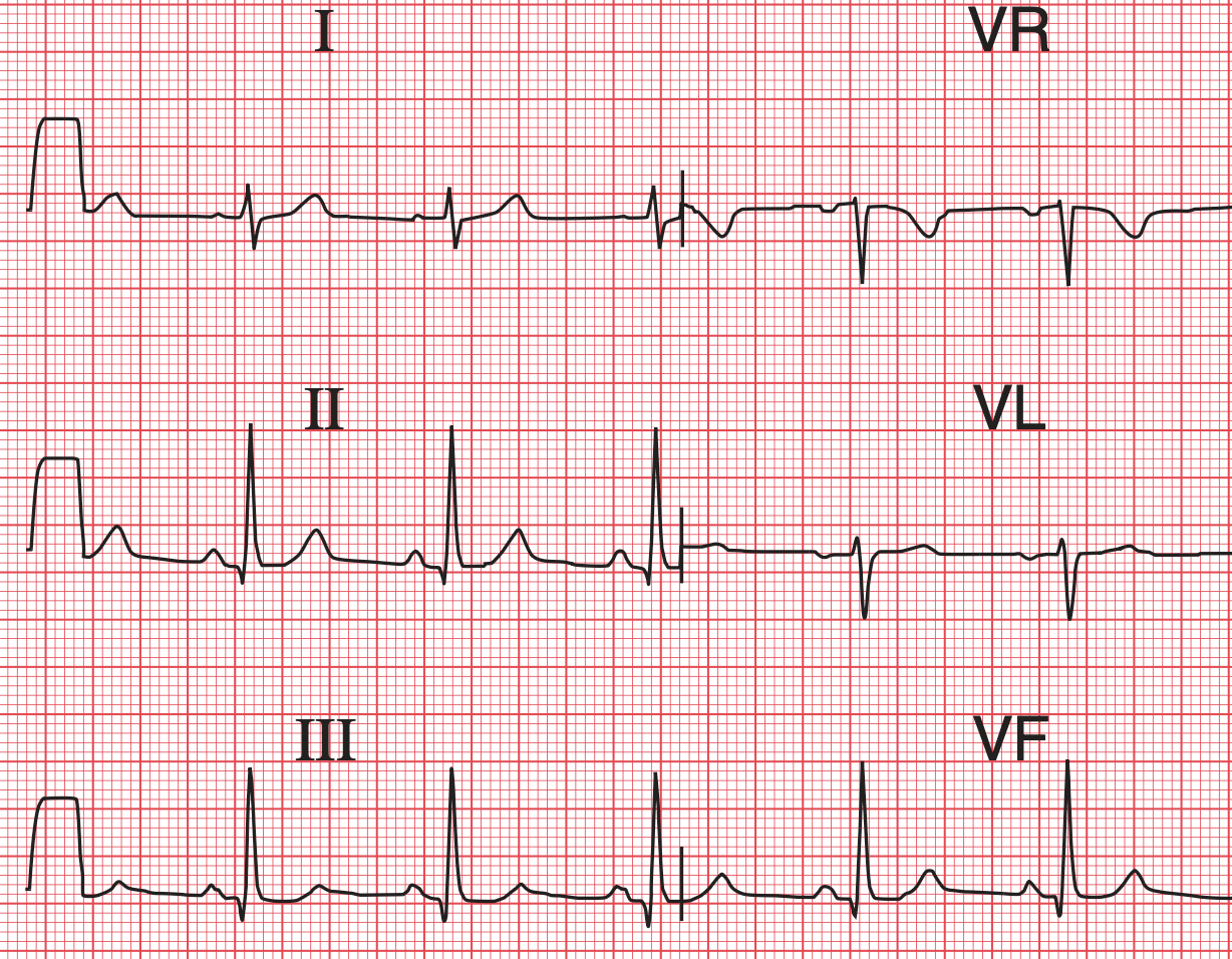
- Right axis deviation
- ECG finding: Lead l negative and aVF positive (Reaching = Right)
 Right axis deviation
- ECG finding: Lead l negative and aVF positive (Reaching = Right)
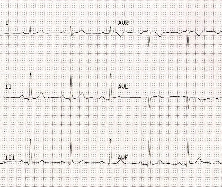
- Left axis deviation
- ECG finding: Lead l positive and aVF negative (Leaving = Left)
 Left axis deviation
- ECG finding: Lead l positive and aVF negative (Leaving = Left)
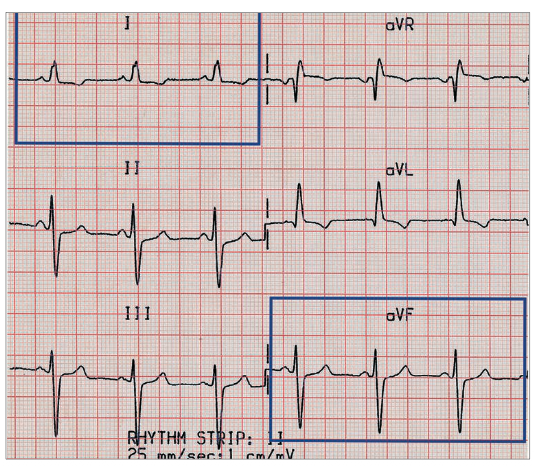
- Extreme axis deviation
- ECG finding: Lead l and aVF being negative
 Extreme axis deviation
- ECG finding: Lead l and aVF being negative
- Hexaxial Reference System
- Find the most equiphasic complex, then move 90 degrees around the Hexaxial Reference System towards the quadrant that you first decided your axis would fall in.
- Normal range -30 to +90
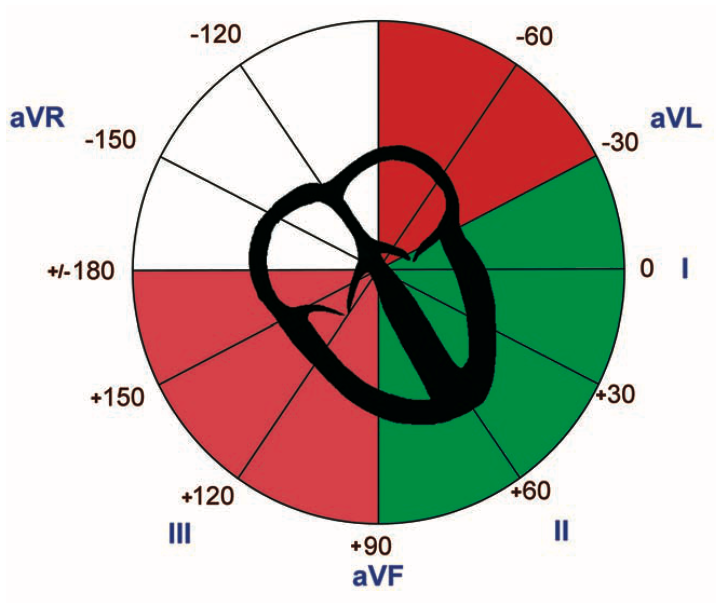 Hexaxial Reference System
Chest Lead
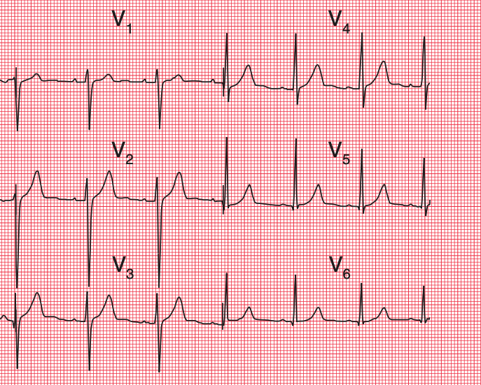 Chest lead ECG
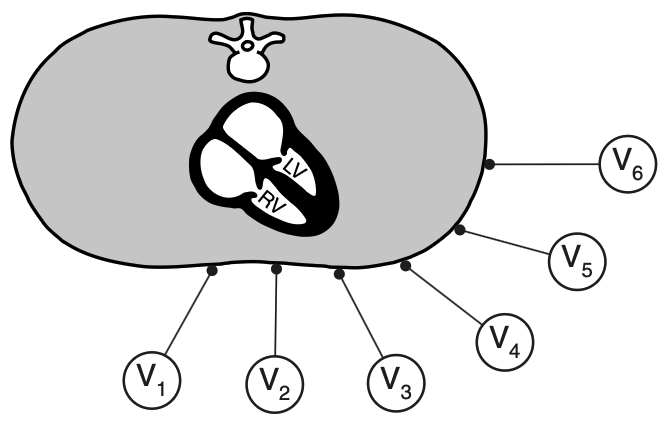 Visual of the chest lead
QRS Complex
- R wave: 1st positive deflection
- Progression V1 - V4, regression V5 - V6
- S wave: negative deflection after R wave
- Regression until V6, absent at V6
- Q wave: 1st negative deflection
- 1st present at V4, biggest at V6
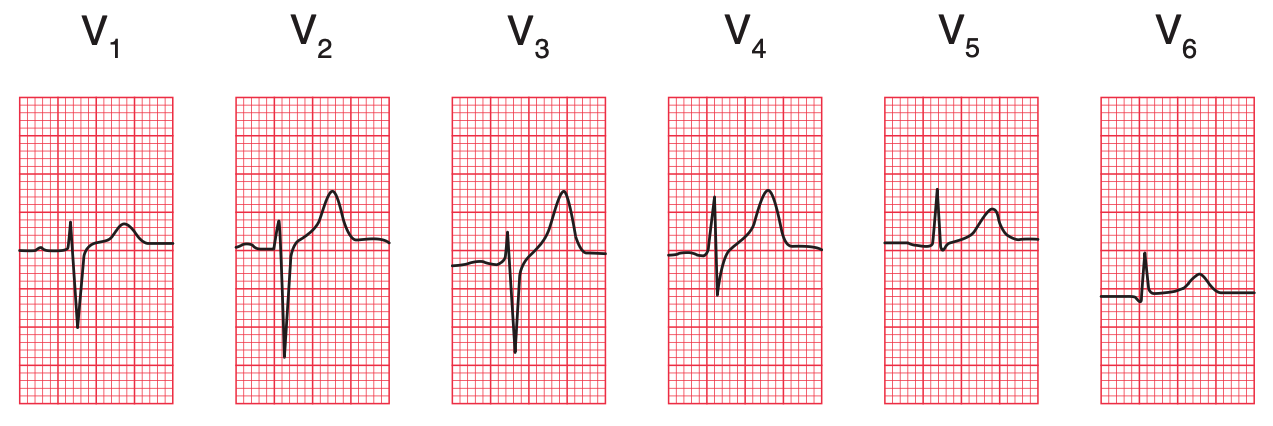 Good R progression with transition at V4, good S regression with total absent at V6, Q wave present at V4-V6
- 1st present at V4, biggest at V6
Heart Injury
- Include ischemia, infarction and necrosis.
- Injury can happen at any part of the heart.
- Locate pathological change at the respective site of ECG.
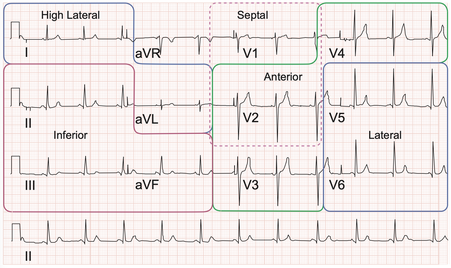 ECG territories
Ischemic heart injury
- ST depression, T inversion and T flattening
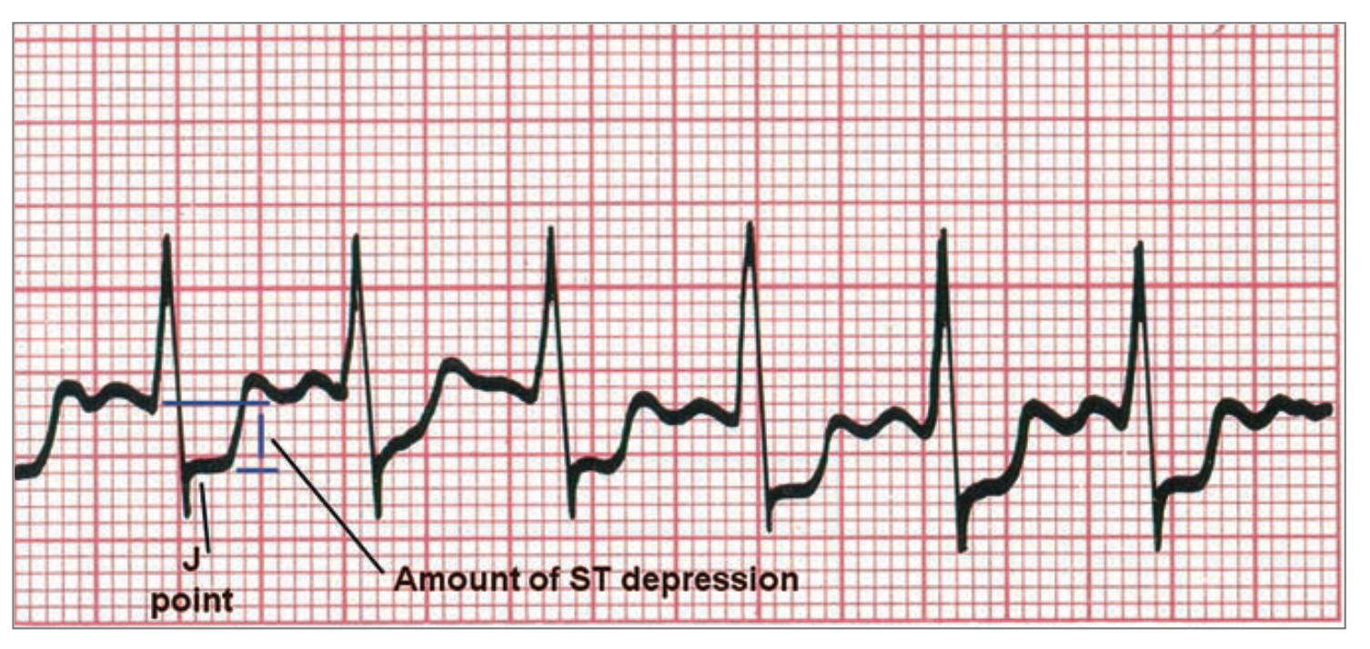 ST depression
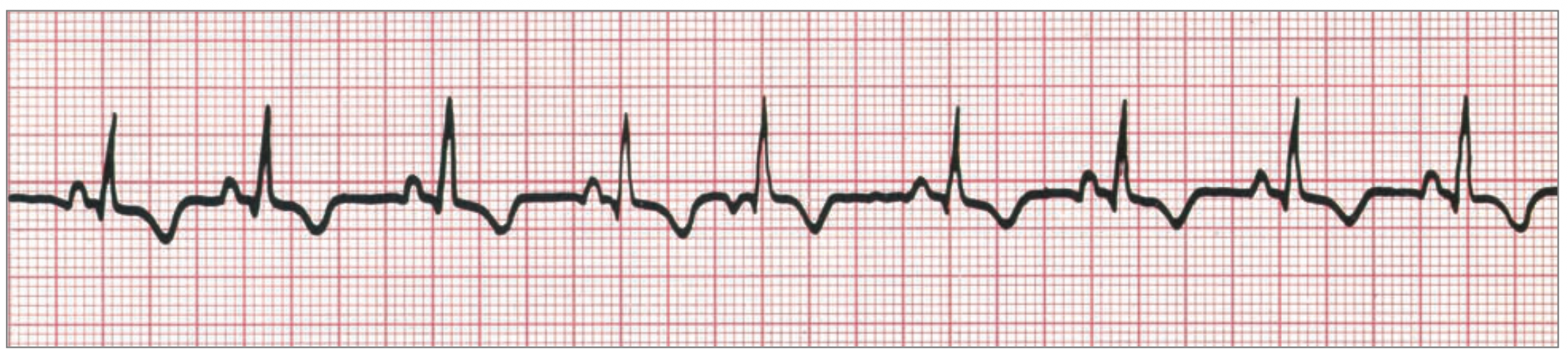 T inversion
Myocardial infarction
- ST elevation
- Reciprocal ST depression at counter site; PAILS
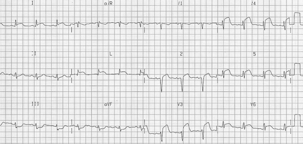 ST elevation at V2-V6, l and aVL, reciprocal change lll and aVF *anterolateral myocardial infarction
Heart necrosis
- Deep Q wave (> 25% depth of R wave)
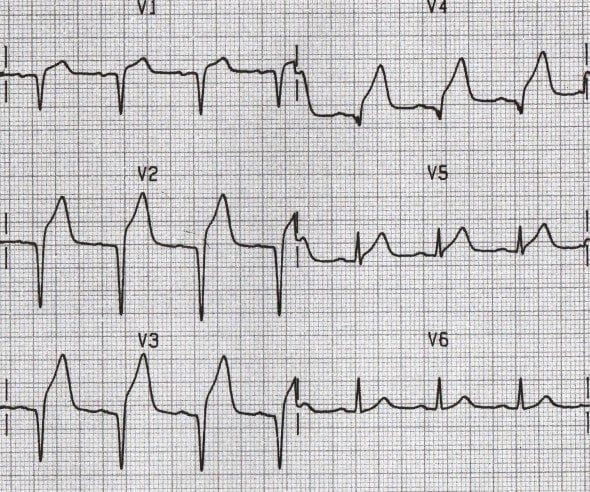 Deep Q wave at V1-V4 with ST elevation *anterior myocardial necrosis with myocardial infarction
Bundle Branch Block
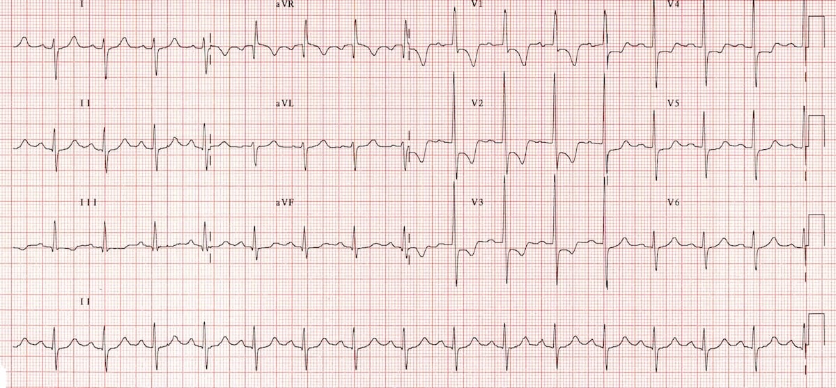
- Right Bundle Branch Block
- ECG finding: V1= RSR pattern (letter M), V6= wide slurred S wave (MaRRoW)
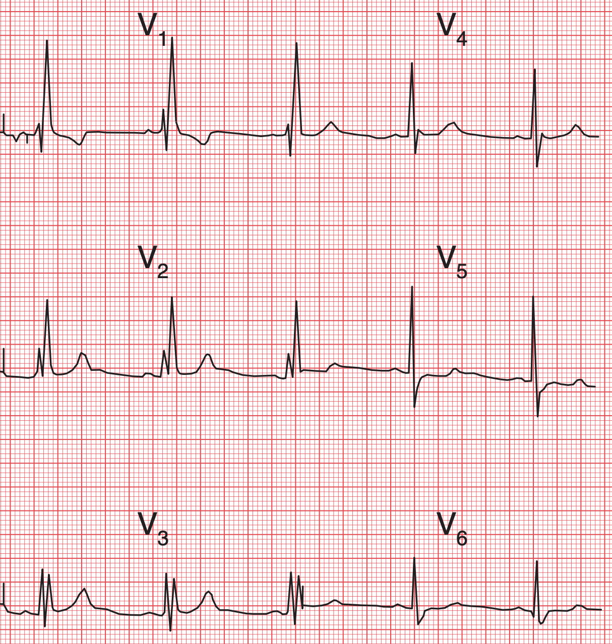 RSR pattern at V1, slurred S wave at V6 *RBBB
- ECG finding: V1= RSR pattern (letter M), V6= wide slurred S wave (MaRRoW)
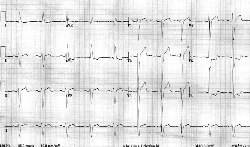
- Left Bundle Branch Block
- ECG finding: V1= rS pattern, V6= M pattern / monophasic R wave (WiLLiaM)
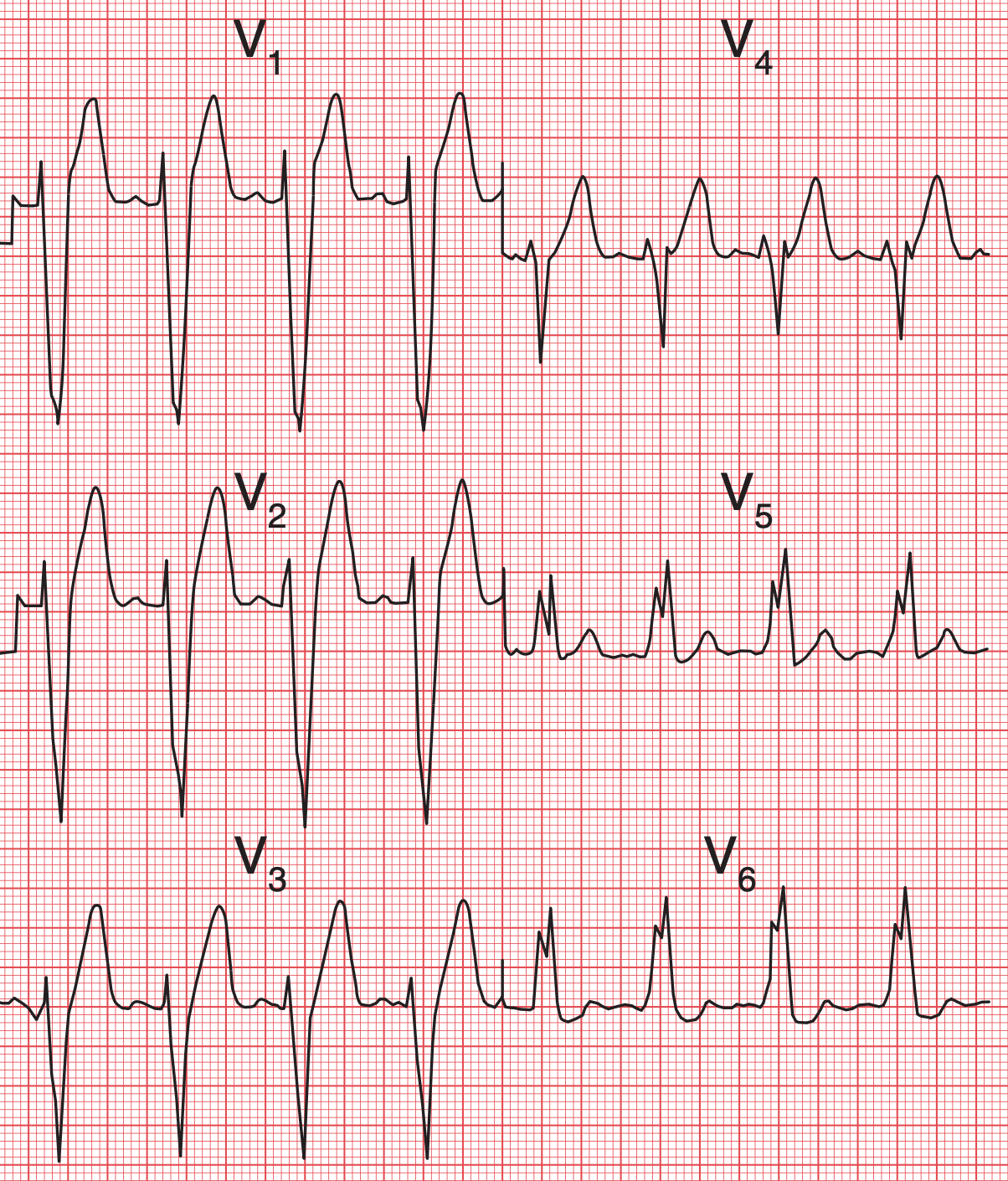 rS pattern at V1, M pattern at V6 * LBBB
- ECG finding: V1= rS pattern, V6= M pattern / monophasic R wave (WiLLiaM)
Chamber Enlargement
- Right atrial enlargement
- ECG finding: Lead ll= P-pulmonale, V1= P-pulmonale
- ECG finding: Lead ll= P-pulmonale, V1= P-pulmonale
- Left atrial enlargement
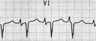
- ECG finding: Lead ll= P-mitrale, V1= P-biphasic
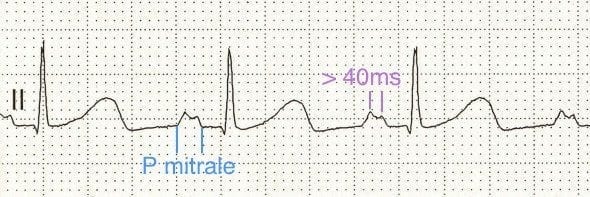
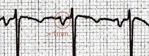
- ECG finding: Lead ll= P-mitrale, V1= P-biphasic
- Right ventricular hypertrophy
- ECG finding: V1= Big R, V5= Big S, Right axis deviation
- ECG finding: V1= Big R, V5= Big S, Right axis deviation
- Left ventricular hypertrophy
- ECG finding: V1= Huge S, V5/V6= Huge R, S + R > 35mm
 Huge S wave at V1, huge R wave at V6, total < 35mm
- ECG finding: V1= Huge S, V5/V6= Huge R, S + R > 35mm
Pos ini dilesenkan di bawah
CC BY 4.0
oleh penulis.